.svg)


Are you looking for notes on human reproduction in Class 12 Biology Chapter 3? Well, you’re in the right place! These notes provide you with information on the male and female reproductive systems, spermatogenesis and oogenesis, the menstrual cycle, fertilisation, pregnancy, parturition and lactation.
These notes from CBSE Class 12 Biology Syllabus aim at providing a simple and examination-orientated understanding of human reproduction, thus being helpful for the CBSE board exams, NEET and last-minute revisions.


Human Reproduction Process
Human beings are sexually reproducing and viviparous organisms. This complex process consists of gamete formation and transfer, followed by fertilization, formation of the zygote and embryogenesis.
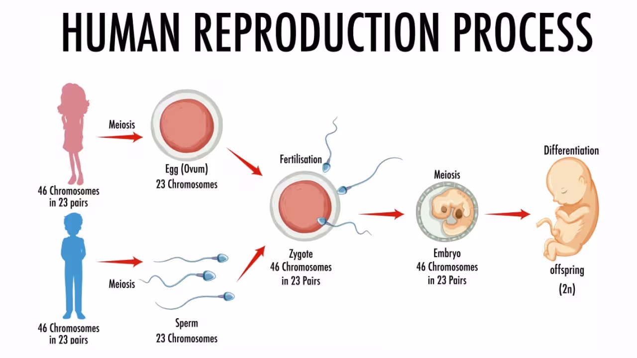
Reproduction ensures the continuity of the species as well as the increase in the number of individuals during favorable conditions. The presence of male and female individuals having different reproductive structures is called sexual dimorphism.
The Male Reproductive System
The male reproductive system is located in the pelvis region and includes a pair of testes, accessory ducts, glands and external genitalia. (Refer to "Figure 2.1a" in NCERT for an overall view of the male reproductive system).
![Untitled.png [apng-to-avif output image]](https://www.educart.co/img-cache/https%3A%2F%2Fcdn.prod.website-files.com%2F5f5cf4627107791c0412287b%2F699d6440c4ece51252ffb5f7_ezgif-4a7e63fd2215140e.avif)
Testes (Testicles)
The testes are located outside the abdominal cavity in a pouch called the scrotum. The scrotum maintains the testes at a temperature 2-2.5°C lower than body temperature, which is necessary for spermatogenesis (sperm formation).
Each testis is oval in shape, about 4-5 cm long and 2-3 cm wide and contains nearly 250 testicular lobules.
Each lobule has 1-3 highly coiled seminiferous tubules, which are the actual sites of sperm production. These tubules contain two important types of cells:
- Sertoli cells, which nourish and support developing sperm
- Spermatogonia, which are diploid germ cells (46 chromosomes) that undergo meiosis to form sperm
Between the seminiferous tubules lies connective tissue containing blood vessels and Leydig cells. Leydig cells secrete androgens (testosterone), which control male sexual development and reproductive functions.
Male Sex Accessory Ducts
The accessory ducts help in storage and transport of sperm. Sperm move through these ducts in a definite sequence.
- Rete testis collects sperm from seminiferous tubules
- Vasa efferentia carry sperm to the epididymis
- Epididymis is a long, coiled tube where sperm mature and are stored
- Vas deferens carries sperm upward, loops over the urinary bladder
- Ejaculatory duct is formed by the vas deferens and seminal vesicle duct and opens into the urethra
- The urethra runs through the penis and serves as a common passage for urine and semen.
Male Accessory Glands
The male accessory glands include paired seminal vesicles, a prostate gland and paired bulbourethral (Cowper’s) glands. Their combined secretions form seminal plasma, which nourishes and protects sperm.
- Seminal Vesicles: They contribute about 60–75% of semen volume. Their secretion contains fructose, prostaglandins, vitamin C, enzymes and proteins that provide energy to sperm.
- Prostate Gland: The prostate secretes a thin, milky, slightly alkaline fluid. This helps neutralize the acidic vaginal environment and improves sperm motility.
- Bulbourethral Glands: Their secretion lubricates the penis and neutralizes residual acidity in the urethra.
- Semen is a mixture of sperm and seminal plasma.
- Penis (External Genitalia): The penis is a cylindrical organ used to deliver sperm into the vagina during intercourse. It is covered by a loose fold of skin called the foreskin (prepuce).
The Female Reproductive System
The female reproductive system is located in the pelvic region and includes ovaries, oviducts, uterus, cervix, vagina, external genitalia and mammary glands.
![image.png [apng-to-avif output image]](https://www.educart.co/img-cache/https%3A%2F%2Fcdn.prod.website-files.com%2F5f5cf4627107791c0412287b%2F699d6441c4ece51252ffb624_ezgif-45d1c3886c5a0be3.avif)
These organs are specialized for ovulation, fertilization, pregnancy, childbirth and nourishment of the newborn.
Ovaries
Ovaries are the primary female sex organs. They produce ova and ovarian hormones like estrogen and progesterone.
Each ovary is about 2-4 cm long and lies on either side of the lower abdomen. Structurally, the ovary has an outer cortex and an inner medulla, covered by a thin epithelium.
The number of ova is fixed before birth. From puberty to menopause, usually one ovum is released every month under hormonal control.
Female Accessory Ducts
These ducts include the oviducts, uterus and vagina.
Oviducts (Fallopian Tubes) are about 10-12 cm long and connect the ovaries to the uterus. Each tube has four regions:
- Infundibulum – funnel-shaped, close to the ovary
- Fimbriae – finger-like projections that collect the ovum
- Ampulla – widest part and site of fertilization
- Isthmus – narrow part joining the uterus
The uterus (womb) is an inverted pear-shaped organ where the embryo develops. It is supported by ligaments.
The uterine wall has three layers:
- Perimetrium: outer thin layer
- Myometrium: thick muscular layer; contracts during childbirth
- Endometrium: inner glandular layer; site of implantation and menstrual changes
Cervix and Vagina
The cervix is the narrow lower part of the uterus. Its opening is called the cervical canal. Along with the vagina, it forms the birth canal.
The vagina connects the cervix to the external genitalia. It acts as:
- passage for menstrual flow
- site of sexual intercourse
- birth canal during delivery
Female External Genitalia
These include the mons pubis, labia majora, labia minora, hymen and clitoris. The mons pubis is a fatty pad covered with pubic hair. The labia majora are fleshy folds surrounding the vaginal opening, while the labia minora lie inside them. The hymen may partially cover the vaginal opening, but it is not a reliable indicator of virginity. The clitoris is a small, sensitive, finger-like structure located above the urethral opening.
Mammary Glands
Mammary glands are modified sweat glands made of glandular and adipose tissue. Each breast has 15-20 lobes and each lobe contains alveoli that produce milk.
Milk flows from alveoli → mammary tubules → mammary ducts → lactiferous ducts, which open at the nipple.
The first milk after childbirth is colostrum. It is yellowish, sticky, rich in antibodies and provides immunity to the newborn.
Gametogenesis
Gametogenesis is the process by which haploid gametes (sperm and ova) are formed in the testes and ovaries.
Spermatogenesis (Sperm formation)
Spermatogenesis occurs in the seminiferous tubules and begins at puberty. It involves mitosis, meiosis and differentiation to form mature sperm cells.
Hormonal Regulation of Spermatogenesis:
- GnRH from hypothalamus stimulates pituitary
- LH acts on Leydig cells to produce testosterone
- FSH acts on Sertoli cells to support spermiogenesis
Spermiogenesis and Spermiation
Spermatids transform into mature spermatozoa through a process called spermiogenesis. During this process, the nucleus condenses, acrosome forms, and tail develops.
After spermiogenesis, sperm heads are embedded in Sertoli cells and are finally released from seminiferous tubules by a process called spermiation.
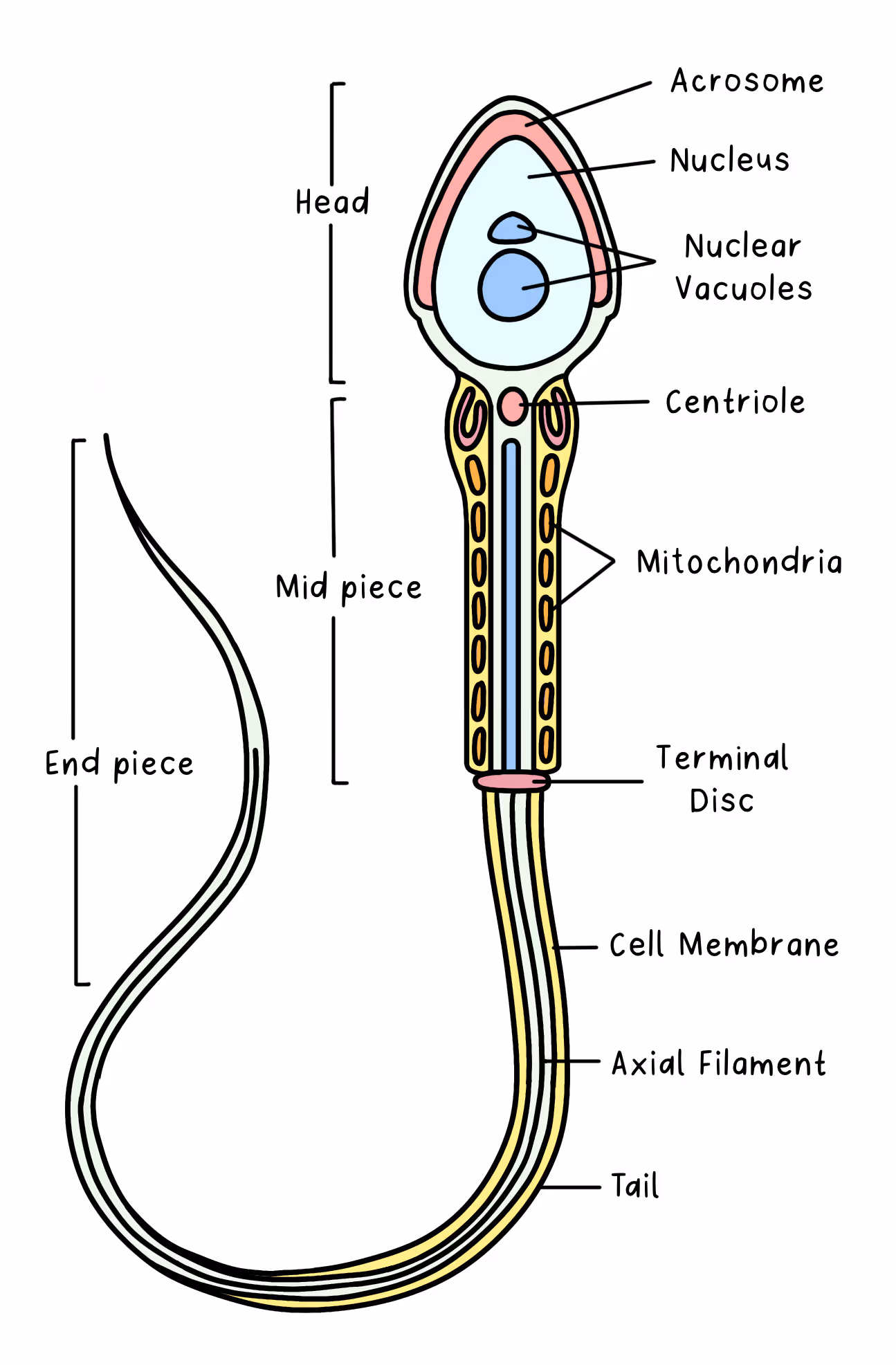
Structure of a Sperm
A sperm has three main parts: head, middle piece and tail. The head contains a haploid nucleus and is capped by an acrosome filled with enzymes that help penetrate the ovum. The middle piece is packed with mitochondria for energy. The tail helps in movement toward the ovum.
For normal fertility, 200-300 million sperm are ejaculated. At least 60% should be normal and 40% should be actively motile.
Oogenesis (Ovum Formation)
Oogenesis is the formation of an ovum from an oogonium and occurs in the ovaries.
It begins before birth and differs greatly from spermatogenesis.
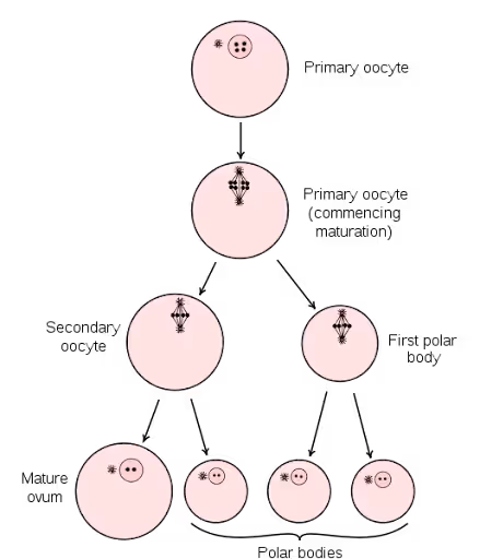
Stages of Oogenesis
- Oogonia form primary oocytes during embryonic life
- Primary oocytes remain arrested in prophase I
- Many follicles degenerate (atresia) before puberty
- Some follicles mature into tertiary follicles
- Meiosis I produces a large haploid secondary oocyte and a small first polar body
- The mature Graafian follicle releases the secondary oocyte during ovulation
- Meiosis II completes only after fertilisation
Menstrual Cycle
The menstrual cycle occurs in female primates. Menarche begins at 10–15 years and menopause occurs around 50 years. The cycle lasts about 28–29 days.
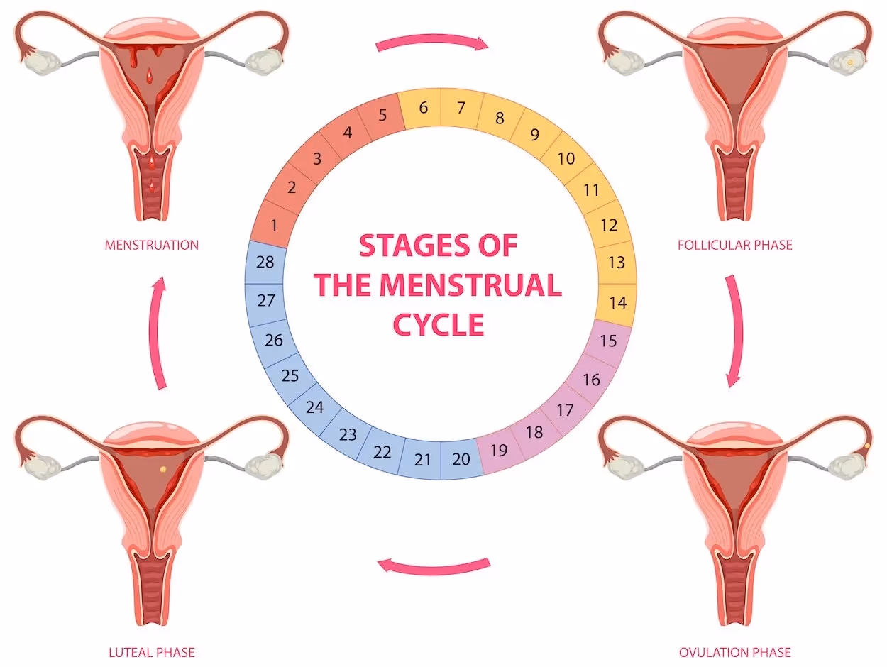
Phases of the Menstrual Cycle
These are the phases of the menstrual cycle:
- Menstrual Phase (Day 1-5): Shedding of the endometrial lining occurs if fertilization does not happen.
- Follicular Phase (Proliferative Phase): FSH and LH stimulate follicle development. Estrogen repairs and thickens the endometrium.
- Ovulatory Phase (Mid-cycle): Around day 14, a sudden LH surge causes ovulation.
- Luteal Phase (Secretory Phase): Corpus luteum secretes progesterone to maintain the endometrium. If fertilization fails, menstruation occurs.
Fertilization and Implantation
Fertilization occurs in the ampullary region of the fallopian tube. Fusion of sperm and ovum forms a diploid zygote.
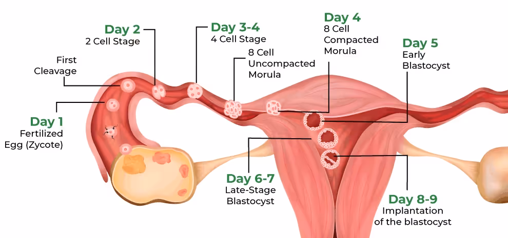
Key steps include acrosomal reaction, prevention of polyspermy, completion of meiosis II and formation of a zygote.
Sex is determined by the father:
- XX → female
- XY → male
Pregnancy and Embryonic Development
Placenta forms from chorionic villi and connects maternal and fetal blood supply.
Functions of the Placenta
- Nutrition and respiration
- Waste removal
- Hormone secretion (hCG, hPL, estrogen, progesterone)
The embryo differentiates into ectoderm, mesoderm and endoderm, which form all body tissues.
Placenta also secretes–hCG, hPL, Estrogens, and Progesterone. During later pregnancy, the ovary secretes relaxin. Maternal levels of prolactin, cortisol and thyroxine also increase to support fetal development.
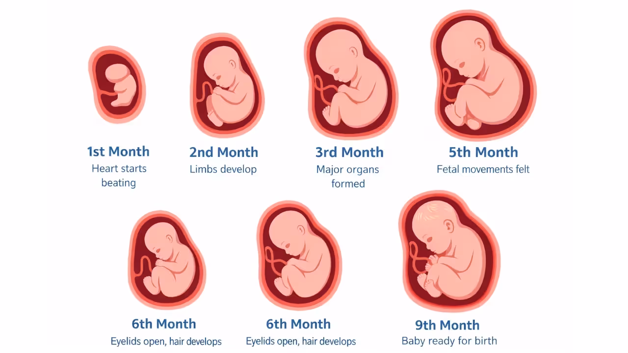
Gestation Period and Fetal Development Milestones
Human pregnancy lasts about 9 months.
- 1st month: Heart starts beating
- 2nd month: Limbs develop
- 12 weeks: Major organs formed
- 5th month: Fetal movements felt
- 24 weeks: Eyelids open, hair develops
- 9 months: Baby ready for birth
Parturition (Childbirth)
Parturition is the process of giving birth to a fully developed fetus at the end of gestation. It's controlled by a complicated neuroendocrine system.
During pregnancy, the fetus and placenta communicate and, together with the maternal pituitary, secrete oxytocin, stimulating weak uterine contractions. This is the fetal ejection reflex. Signals from the fully developed foetus and placenta initiate mild uterine contractions called the foetal ejection reflex.
This triggers the release of oxytocin from the maternal pituitary gland, which causes stronger uterine contractions leading to childbirth.
Lactation
Lactation is the secretion of milk by the mammary glands that are differentiated during pregnancy, after giving birth. The milk secreted in the first few days is colostrum, which is a sticky, yellow fluid with high antibody (immunoglobulin) concentrations, which gives the newborn innate immunity.
The colostrum is also low in fats while high in proteins, which makes it easier to support growth. During this period, breastfeeding is very important for the newborn.
Conclusion
Human reproduction may look complicated at first, but once you understand the sequence - gamete formation, fertilisation, implantation and development - everything connects logically.
If you focus on diagrams, hormonal control and key stages like ovulation, fertilisation and parturition, this chapter becomes scoring and easy. That’s a wrap on Human Reproduction! Revise the diagrams once more and you’re exam-ready.
FAQs
Q1. What are the main female external genitalia?
Ans: The female external genitalia include the mons pubis, labia majora, labia minora, hymen and clitoris. They protect internal organs and aid in sexual function.
Q2. What are the functions of the fallopian tubes (oviducts)?
Ans: Fallopian tubes (10–12 cm) transport the ovum from ovary to uterus. Fertilisation usually occurs in the ampulla.
Q3. What is spermatogenesis and where does it occur?
Ans: Spermatogenesis is the formation of sperm from spermatogonia. It occurs in the seminiferous tubules of the testes and is regulated by FSH, LH and testosterone.
Q4. How does fertilisation occur and how is the sex of a baby determined?
Ans: Fertilisation is the fusion of sperm and ovum in the fallopian tube, forming a zygote. Ovum carries X; sperm carries X or Y. XX = female, XY = male.
Q5. Why is the scrotum located outside the body in males?
Ans. The scrotum is outside the body to maintain the testes at a temperature about 2-2.5°C lower than body temperature, which is essential for spermatogenesis (sperm production).