.svg)

Have you ever thought about how the food you eat turns into energy? That’s where the alimentary canal comes in - it’s basically your body’s own food highway. From the moment food enters your mouth to when waste leaves your body, this amazing system makes sure every bite is used efficiently.
It includes important parts like the stomach, small intestine, and large intestine, plus several other organs and enzymes that work together to keep digestion smooth.
In this blog, we will let you know what the alimentary canal is, its main parts, how it works, and why it’s so important all in a student-friendly way.
What is the Alimentary Canal?
The alimentary canal is basically the main pathway through which food travels in our body. It’s a long, muscular tube that starts from the mouth and ends at the anus, designed to break food down into nutrients your body can use and then remove waste you don’t need.
Think of it like a well-organized food-processing line -where each part of the canal has its own job. The canal isn’t just about digestion; it’s about absorbing nutrients, sending them into your bloodstream, and making sure waste leaves your body smoothly.
Here’s what makes the alimentary canal special:
- Length: In humans, it’s about 8 meters long, making it a super efficient system.
- Purpose: To digest food, absorb nutrients, and get rid of waste.
- Key organs involved: Oesophagus, stomach, small intestine, large intestine, and anus.
- Teamwork with other organs: It works with stomach enzymes and organs of the alimentary canal like the liver, pancreas, and salivary glands to make digestion possible.
In short, the alimentary canal is your body’s digestive superhighway - moving food along, breaking it down, and keeping you nourished.


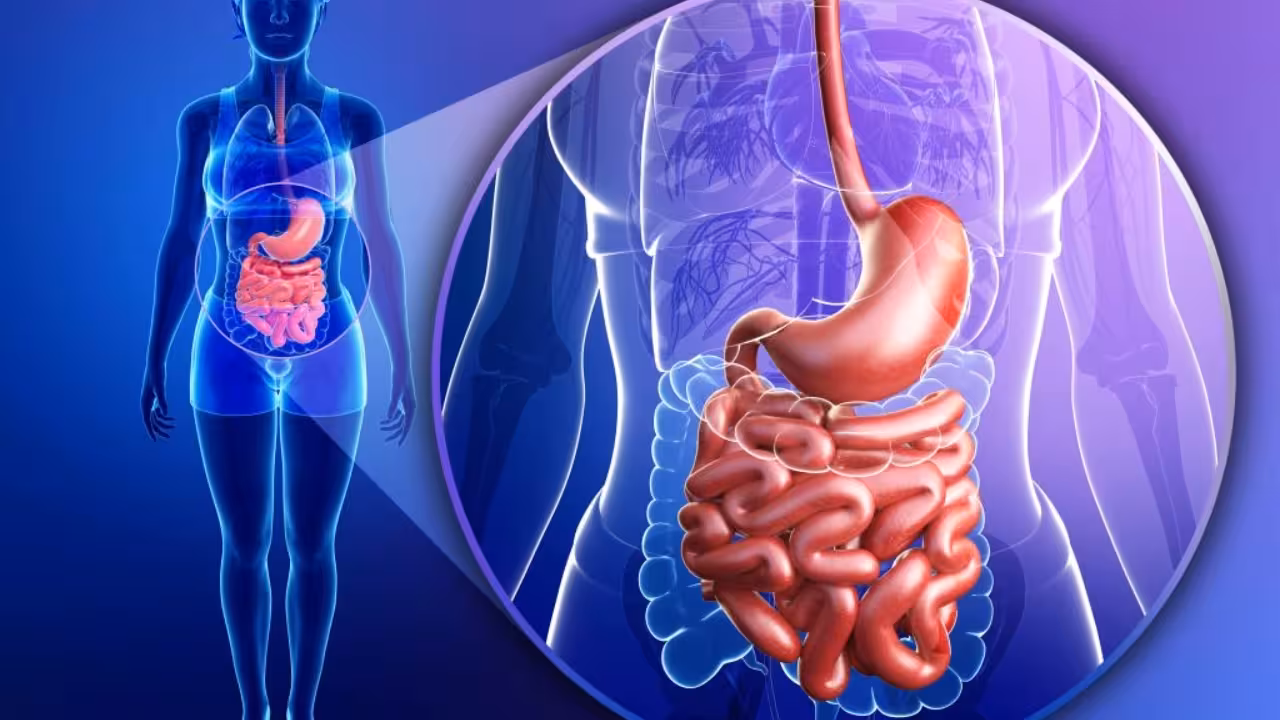
Major Parts of the Alimentary Canal
Think of the alimentary canal as your body’s ultimate food processing line. It starts the moment you take a bite and ends when your body gets rid of waste. Each part has a special job to make sure food turns into nutrients your body can actually use. Let’s break it down.
.avif)
1. Mouth and Buccal Cavity
This is where digestion kicks off. Food enters here, gets chewed by teeth, and mixed with saliva. Saliva isn’t just wetness - it contains enzymes like amylase that start breaking down starch. The tongue helps mix the food and push it toward the next stage.
2. Pharynx
The pharynx is basically a crossroads. It connects your mouth to the oesophagus and makes sure food goes the right way while keeping it away from the airway.
3. Oesophagus
The oesophagus is like a muscular food chute, about 25 cm long. It uses a squeezing motion called peristalsis to push food toward the stomach. No gravity needed - it works even if you’re upside down!
4. Stomach
The stomach is where food gets a real makeover. It’s a J-shaped muscular sac that stores and churns food with gastric juices. These juices contain stomach enzymes like pepsin that break proteins into smaller pieces. The result is a semi-liquid paste called chyme, ready for the small intestine.
5. Small Intestine
The small intestine is where most digestion and absorption happens. It’s the longest part of the alimentary canal, measuring around 6 - 7 meters! It has three parts:
- Duodenum: mixes chyme with bile and pancreatic juices.
- Jejunum: absorbs nutrients like sugars and amino acids.
- Ileum: absorbs remaining nutrients and bile salts.
This is where the magic of the small intestine function happens - turning digested food into energy and nutrients.
6. Large Intestine
The large intestine (about 1.5 meters long) is mainly about water absorption and turning food into solid waste. It includes: cecum, colon, rectum, and anus. Its job is essential: keeping your body hydrated and making stools (function large intestines).
7. Anus
Finally, the anus is the exit point of the alimentary canal. Controlled by muscles, it makes sure waste leaves your body at the right time.
Accessory Organs of Digestion
Okay, so the alimentary canal does most of the digestion work - but it doesn’t do it alone. Some other organs help by releasing juices, enzymes, and bile that make food breakdown easier. These are called accessory organs of digestion. Let’s meet them:
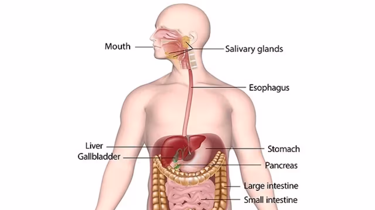
1. Salivary Glands
- There are three pairs - parotid, submandibular, and sublingual glands.
- They secrete saliva, which contains salivary amylase (the enzyme that starts digesting starch into sugar).
- Basically, they make food soft and chemical digestion starts right in your mouth.
2. Liver
- The largest gland in your body and a real multitasker.
- It produces bile, which doesn’t digest food directly but helps break down fats into tiny droplets (a process called emulsification).
- Bile is stored in the gallbladder until needed.
- The liver also filters toxins and manages nutrients absorbed from food - total boss of metabolism.
3. Gallbladder
- A small, pear-shaped pouch sitting under the liver.
- Its job: store and concentrate bile before releasing it into the small intestine.
- Helps digest fatty foods - like oils, butter, and cheese.
4. Pancreas
- The pancreas acts as both a digestive and endocrine organ.
- It secretes pancreatic juice, rich in enzymes like:
- Trypsin (for proteins)
- Amylase (for starch)
- Lipase (for fats)
- These enzymes mix with food in the small intestine and complete the digestion process.
Layers of the Alimentary Canal
When you cut across the alimentary canal like the stomach or intestines, you’ll notice it’s not just a simple tube - it’s made of four main layers, each with its own special job. Let’s break it down like an anatomy pro.
.avif)
1. Mucosa – The Inner Working Layer
- It’s the innermost lining that comes in direct contact with food.
- Has glands that secrete digestive juices, mucus, and enzymes.
- In the stomach, it releases acid and stomach enzymes like pepsin.
- In the small intestine, it forms tiny folds called villi that absorb nutrients.
Think of mucosa as the “active surface” where digestion and absorption actually happen.
2. Submucosa – The Support System
- Lies right under the mucosa.
- Made up of connective tissue, blood vessels, and nerves.
- Supplies nutrients to the mucosa and helps control its secretions.
- Contains nerve networks called the Meissner’s plexus (important for coordination).
3. Muscularis – The Movement Machine
Let's look at how your gut muscles keep it moving - this is where the muscularis layer comes into play.
Without this muscular action, food would just sit still in your bowel - and digestion wouldn’t work at all.
4. Serosa – The Outer Protective Layer
- The outermost layer, made of thin connective tissue.
- Protects the canal and reduces friction as organs move.
- It’s like the “wrapper” that keeps your stomach and intestines safe and smooth inside your abdomen.
The four layers of the alimentary canal - mucosa, submucosa, muscularis, and serosa - work together to digest, absorb, and move food efficiently through the small intestine and large intestine.
Digestive Enzymes and Secretions
The alimentary canal is full of chemical action - every part releases specific digestive enzymes and secretions that help break food into absorbable nutrients. Let’s go through them step-by-step.
.avif)
1. Mouth & Salivary Glands
Let’s see how each organ adds its own enzymes and secretions to make digestion happen.
Remember: Digestion starts in the mouth - saliva softens food and starch breakdown begins here.
2. Stomach
The stomach’s gastric juice (with pepsin, rennin & HCl) digests proteins and turns food into chyme.
The stomach is basically a muscular acid chamber - it mixes food into a semi-liquid called chyme.
3. Small Intestine
Pancreatic, intestinal juices & bile complete digestion - breaking proteins, fats, and sugars for absorption.
Reminder: Most digestion actually finishes here - the small intestine is the main site for enzyme action and nutrient absorption.
4. Large Intestine
- No major digestive enzymes are secreted here.
- Absorbs water, minerals, and vitamins produced by gut bacteria.
- Helps form solid waste (feces).
Every step in digestion depends on these enzymes and secretions - from salivary amylase in the mouth to pancreatic lipase in the small bowel intestine. Together, they ensure food is broken down, absorbed, and ready to fuel your body.
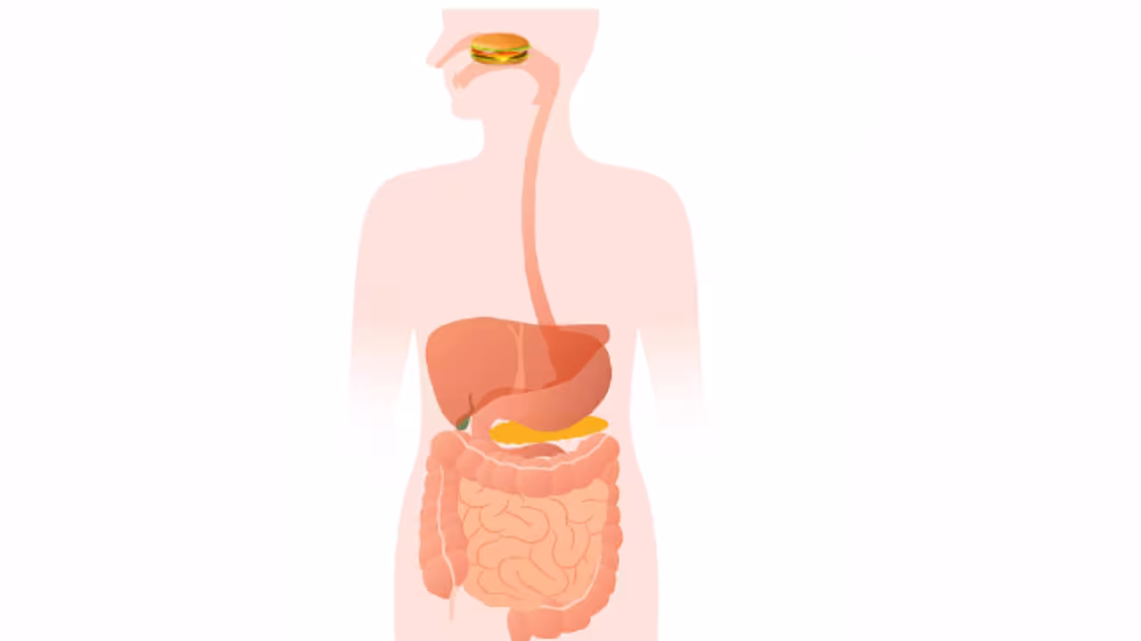
Peristalsis and Movement of Food
Once food enters your alimentary canal, it doesn’t just slide down by gravity - it’s actually pushed and mixed by a rhythmic muscle movement called peristalsis. Think of it as the canal’s own wave system: smooth muscles contract and relax in sequence to move food forward through your oesophagus, stomach, small intestine, and large intestine.
Step-by-Step Movement Along the Canal
1. Mouth to Esophagus:
- After chewing, food turns into a soft ball called a bolus.
- It’s pushed into the oesophagus, where peristaltic waves start - squeezing food down toward the stomach.
2. Esophagus to Stomach:
- These muscular contractions continue until the food reaches the stomach.
- The lower esophageal sphincter opens briefly to let food in, then closes to prevent acid reflux.
3. Inside the Stomach:
- The stomach muscles churn food with gastric juice to form chyme.
- Peristalsis here helps mix food and slowly release it into the small intestine.
4. In the Small Intestine:
- Gentle, wave-like contractions move chyme through the duodenum, jejunum, and ileum.
- These movements mix food with bile, pancreatic juice, and intestinal enzymes, ensuring complete digestion and nutrient absorption.
5. In the Large Intestine:
- The leftover material is slowly pushed along the bowel large intestine.
- Water and minerals are absorbed while feces are formed and moved toward the rectum.
Why Peristalsis Matters?
- Keeps food moving one way only - mouth → anus.
- Helps mix food with digestive secretions.
- Prevents blockages or backflow of stomach acid.
- Essential for normal digestion and nutrient absorption.
Peristalsis is the reason food doesn’t just sit still - it’s a continuous, coordinated muscle movement that keeps everything flowing smoothly through your stomach, small intestine, and large intestine till digestion is complete.
Blood Supply and Innervation of the Alimentary Canal
Your alimentary canal works nonstop - digesting, absorbing, and pushing food along. To keep up with this, it needs a steady blood supply and nerve control. Think of it like a power-plus-control system: blood provides fuel and oxygen, while nerves coordinate all movements.
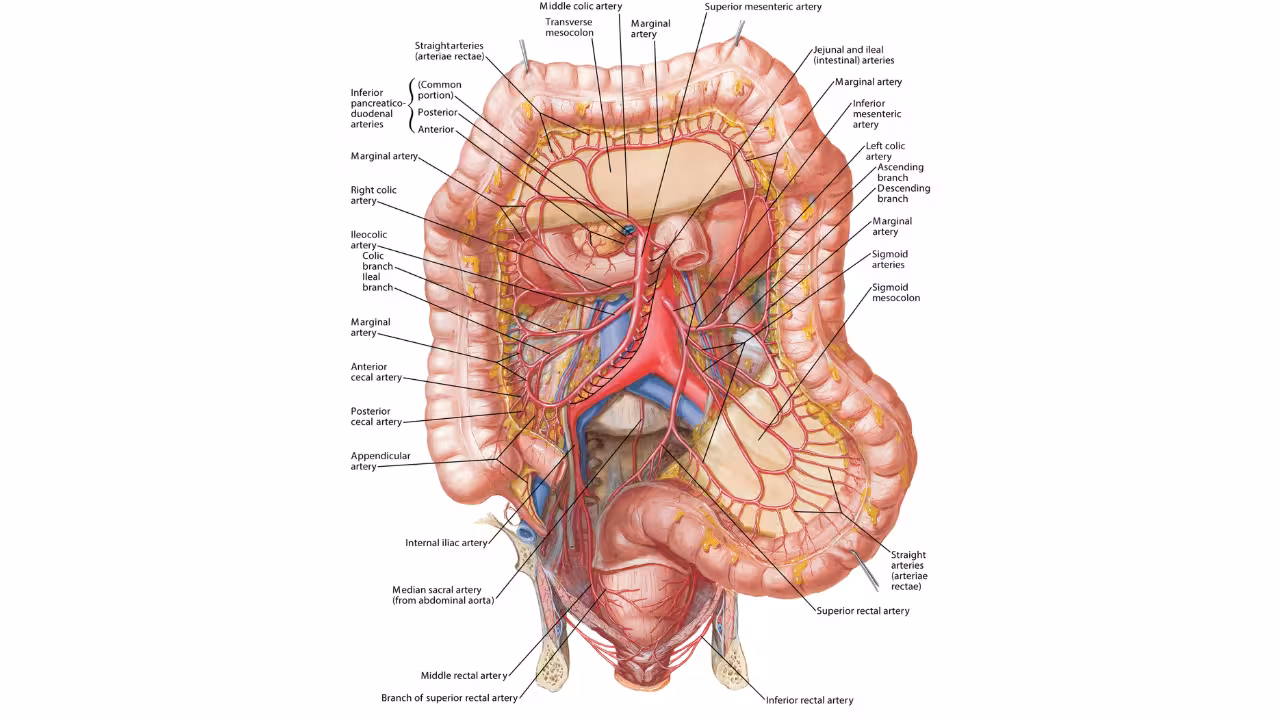
Blood Supply – The Fuel System
Each part of the canal gets its blood from major branches of the aorta:
After digestion, blood from the stomach and intestines doesn’t go straight back to the heart - it first passes through the hepatic portal vein to the liver, where nutrients are processed and toxins are filtered. This is called the hepatic portal circulation.
Nerve Supply – The Control System
Your digestive tract also has its own nerve network known as the enteric nervous system - often called the brain of the gut.
- Parasympathetic nerves (vagus nerve): Stimulate digestion - increase peristalsis, secretion of enzymes, and blood flow.
- Sympathetic nerves: Slow everything down - reduce movement and secretion during stress or danger.
- Enteric plexuses (Auerbach’s & Meissner’s): Local nerve networks that coordinate muscle contractions and gland activity directly inside the gut walls.
The blood supply powers the digestive system, while the nerve supply keeps everything coordinated. Together, they ensure smooth and efficient digestion from start to finish.
Structural Differences in Herbivores vs Carnivores
When it comes to the alimentary canal, not every animal is built the same. Herbivores (plant-eaters) and carnivores (meat-eaters) have different digestive system designs because their diets demand different ways of processing food. Let’s break this down clearly:
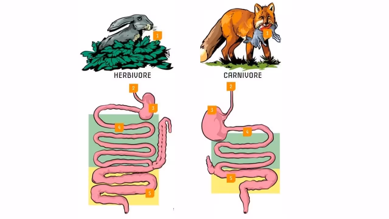
Herbivores (Plant-Eaters)
- Longer alimentary canal: plant matter is harder to digest, so they have a longer small intestine and large intestine to allow more time for digestion.
- Large caecum: important for fermenting cellulose found in plants with the help of gut bacteria.
- Flat teeth with ridges: ideal for grinding plant fibers.
- Examples: Cows, horses, sheep.
Carnivores (Meat-Eaters)
- Shorter alimentary canal: meat is easier to digest, so they don’t need a long intestine.
- Small caecum: less need for cellulose fermentation.
- Sharp, pointed teeth: designed to tear meat.
- Strong stomach acids: to quickly break down proteins and kill bacteria.
- Examples: Lions, tigers, wolves.
The differences in stomach, small intestine, and large intestine anatomy are perfect examples of how the body adapts to diet. It’s basically nature’s engineering at work - designing organs according to what you eat.
Common Disorders Related to Alimentary Canal Anatomy
The alimentary canal is vital for digestion, but like any system in the body, it can face problems. Here are some common disorders related to its anatomy and functioning:
.avif)
- Gastritis
Inflammation of the stomach lining, often caused by infection, excessive alcohol, or spicy food. It affects the stomach’s ability to produce enzymes and acids properly.
- Acid Reflux (GERD)
When stomach acid flows back into the oesophagus, causing heartburn and irritation. This can happen if the lower oesophageal sphincter weakens.
- Constipation
Slow movement of waste through the large intestine (bowel large intestine) can lead to difficulty in bowel movements. This may happen due to low fiber intake, dehydration, or weak intestinal muscles.
- Diarrhoea
Rapid movement of food through the small bowel intestine reduces nutrient and water absorption, causing loose stools. Often caused by infection or digestive enzyme problems.
- Peptic Ulcers
Sores in the lining of the stomach or duodenum caused by excess acid or bacterial infection. They can cause pain and digestive discomfort.
- Irritable Bowel Syndrome (IBS)
A functional disorder affecting the large intestine, causing cramping, pain, bloating, and altered bowel habits without visible damage.
- Celiac Disease
An autoimmune disorder where gluten damages the lining of the small intestine, reducing nutrient absorption and causing digestive distress.
Knowing these disorders makes it easier to see how the alimentary canal works. For example, if the small intestine isn’t working well, your body can’t absorb nutrients properly. If the large intestine has a problem, water absorption and stool formation get affected.

Frequently Asked Questions
Q1. What is the alimentary canal and why is it important?
Ans. Think of the alimentary canal as your body’s food highway - a long tube that runs from your mouth all the way to your anus. It’s where food travels, gets broken down, nutrients get absorbed, and waste is pushed out.
Every part has a role, from chewing in the mouth to absorbing nutrients in the intestines. Without it, your body wouldn’t get the fuel it needs to work.
Key Points to Know:
- Purpose: Digests food, absorbs nutrients, removes waste.
- Main parts: Mouth, oesophagus, stomach, small intestine, large intestine.
- Importance: Essential for energy, growth, and health.
Q2. What are the main parts of the alimentary canal?
Ans. The alimentary canal is a chain of organs that food passes through, each with its own job. It starts at the mouth, goes down the oesophagus, into the stomach, passes through the small intestine, moves to the large intestine, and ends at the anus. Each section helps process food step-by-step so the body can get what it needs.
Quick Breakdown:
- Mouth: Chews and mixes food.
- Oesophagus: Moves food to the stomach.
- Stomach: Breaks down proteins and churns food.
- Small intestine: Absorbs nutrients.
- Large intestine: Absorbs water and forms waste.
Q3. What is the function of the alimentary canal in the human body?
Ans. The alimentary canal is basically your body’s digestion system in action. It breaks down food into nutrients your body can use, absorbs those nutrients, and gets rid of the rest as waste. Without it, you wouldn’t be able to extract energy or build new cells.
Function Highlights:
- Breaks down food mechanically and chemically.
- Absorbs nutrients into the bloodstream.
- Removes leftover waste effectively.
Q4. How does food move through the alimentary canal?
Ans. Food moves along your alimentary canal thanks to something called peristalsis - wave-like muscle contractions that push food forward. This movement happens automatically and keeps digestion going without you even thinking about it.
Key Facts:
- Peristalsis happens in the oesophagus, stomach, and intestines.
- Helps mix food with digestive juices.
- Ensures smooth movement of food through the system.
Q5. What is the difference between the small and large intestine?
Ans. Both the small and large intestine are part of the alimentary canal, but they have different roles. The small intestine is the nutrient absorber - it takes food nutrients into the blood. The large intestine mainly absorbs water and prepares waste for removal.
Key Differences:
- Small intestine: 6–7 meters long, absorbs nutrients, lined with villi.
- Large intestine: 1.5 meters long, absorbs water, forms stool, no villi.
- Main role difference: digestion vs waste formation.
Q6. How long is the human alimentary canal?
Ans. On average, the human alimentary canal is about 9 meters long - that’s roughly the length of a small car! Most of it comes from the small intestine, which is long to give your body enough time to digest food and absorb nutrients properly.
Quick Facts:
- Small intestine: about 6–7 meters.
- Large intestine: about 1.5 meters.
- Length helps in complete digestion and absorption.
Q7. What organs are considered accessory to the alimentary canal?
Ans. Accessory organs don’t carry food, but they are key to digestion. They include:
- Salivary glands: produce saliva and enzymes.
- Liver: makes bile to digest fats.
- Gallbladder: stores and releases bile.
- Pancreas: produces digestive enzymes and hormones.
Key Points:These organs release substances that help break down food and absorb nutrients effectively.
Q8. What role does the stomach play in digestion?
Ans. The stomach is like your food mixer - it churns food and mixes it with gastric juices to form a semi-liquid substance called chyme. It produces stomach enzymes and acid to start protein digestion before sending the food to the small intestine.
Stomach Highlights:
- Mechanical digestion: mixing food.
- Chemical digestion: enzymes breaking proteins.
- Controls release of chyme to the small intestine.
Q9. What are common diseases of the alimentary canal?
Ans. Problems in the alimentary canal can mess with digestion and health. Some common disorders are:
- Acid reflux/GERD: stomach acid flows back into the oesophagus.
- Peptic ulcers: sores in the stomach lining.
- Irritable bowel syndrome (IBS): irregular bowel habits.
- Crohn’s disease: inflammation of the canal.
Key Notes:These conditions show why a healthy alimentary canal is vital for digestion and nutrient absorption.
Q10. What is peristalsis and why is it important?
Ans. Peristalsis is the natural, wave-like muscle movement that pushes food through your alimentary canal. It keeps digestion going smoothly without extra effort from you. Without it, food would just sit still and digestion wouldn’t happen properly.
Key Highlights:
- Happens in the oesophagus, stomach, and intestines.
- Helps mix food with digestive juices.
- Ensures steady movement of food to absorb nutrients efficiently.

Table of Content
Further Study